What is patellar luxation?
Patellar luxation (meaning out of place) is a condition in which the kneecap (patellar) moves (or dislocates) out of the trochlear groove, and moves to the inside (medial) or outside (lateral) of the knee joint (known as the stifle joint in cats). Medial luxation is more common than lateral. Unresolved patellar luxation can lead to arthritis.
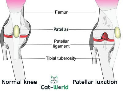
The patellar is what most of us know as the knee bone; it is a small circular bone that sits in front of the knee joint, which consists of the femur in the upper leg and the tibia and fibula in the lower leg. It sits in a vertical groove at the lower end of the femur known as the trochlear groove.
As the hind leg moves, the patellar slides up and down the groove. Buried by the patellar tendon, a strong, flat tendon that attaches from the bottom of the quadriceps muscle of the thigh, over the patellar and attaches to the tibial crest, a bony prominence on the shin bone in the lower leg.
The patellar is a sesamoid bone, the function of which is to act as a pulley, by allowing the tendon to sit further away from the joint, which helps increase movement and strength as the joint straightens and bends. The patellar also helps to protect the knee joint and keep the tendon in place.
The condition can occur in cats of all ages and breeds, although there appears to be a higher incidence in Abyssinians, Bengals, Devon Rexes, British Shorthairs, Siamese, Maine Coons, and Persians.
Causes
There are several possible causes of patellar luxation cats.
- Physical trauma – Traumatic luxations are more painful. They frequently run concurrently with other physical injuries and can affect one joint or both.
- Congenital (present at birth) – This type of patellar luxation tends to be bilateral, affecting both knees. Most often the result of abnormal bone development, as well as a shallow trochlear groove that often slopes towards the side of the luxation.
The patellar can slip out of place if;
- The trochlear groove is too shallow
- The cat has bowed bones
- The tendon attachment is off centre
Symptoms
Cats with mild patellar luxation may not display any signs, particularly with congenital patellar luxation.
- Sudden lameness (traumatic) or intermittent lameness affecting one or both of the hind legs
- As the cat walks, he may skip or hop on the affected leg, or frequently pull the rear leg up towards the body
- Reluctance to jump and climb
- Stiffness in the affected limb(s)
- Cats with medial patellar luxation may walk with a bowlegged gait
Diagnosis
In some cases, patellar luxation may be an incidental finding during a routine physical examination.
The veterinarian will perform a thorough evaluation and obtain a medical history from you. Which will include a lameness examination to evaluate and grade the severity of the patellar luxation.
X-ray images taken from different angles to view the patellar and the depth of the trochlear groove will confirm the diagnosis. The veterinarian will also look for signs of arthritis in the affected knee as a result of the patellar luxation.
Patellar luxation grading:
| Grade 0: | Normal patellar. |
| Grade 1: | The patellar luxates with manual pressure upon full extension of the stifle joint but easily returns to the trochlear groove when released. |
| Grade 2: | The patellar luxates with manual pressure or spontaneously luxate upon flexion of the stifle joint and remains luxated until it is repositioned in the trochlear groove manually. |
| Grade 3: | The patellar is permanently luxated, it can be manually returned to the trochlear groove on extension but re-luxates when released. |
| Grade 4: | The patellar is permanently luxated and cannot be re-positioned in the trochlear groove. The trochlear groove may be shallow or completely absent. |
Treatment
Grades 1 and 2 generally require no treatment; some cats manage to pop the patellar back into place themselves by extending the affected leg. It is advisable to ensure your cat’s weight doesn’t creep up as excess weight places additional pressure on all of the cat’s joints.
Grades 3 and 4 require surgery, preferably before the onset of osteoarthritis. There are several surgical treatments for patellar luxation, which depend on the type of abnormality present.
- Tibial crest transposition: Reposition the attachment of the tendon at the tibial crest to re-align the patellar with the trochlear groove.
- Trochleoplasty: Deepening and/or widening of the trochlear groove.
- Lateral or medial imbrication: It is common for the soft tissue surrounding the patellar to stretch over time. Tightening the tissue during surgery will help to hold the patellar in place.
- Osteotomy: Some grade 4 cases may require surgery to realign the tibia or fibula; this surgery involves cutting the femur and re-aligning it with clamps.
The success rate of this surgery is usually very high, particularly if caught in time before arthritis has developed.
Home care
Antibiotics, painkillers, and anti-inflammatories will be provided upon discharge. Administer all medications as prescribed.
Keep a close eye on the surgical site for signs of infection which includes swelling, redness, and oozing, if you notice any of these, see your veterinarian immediately.
Your veterinarian may recommend gentle exercises for you to perform on your cat during his recovery to help restore his normal range of motion. Follow as instructed as rehabilitation is an important part of recovery.
Confine to a crate or room until recovered, which takes two weeks. Avoid excessive activity and jumping for three months; keep the cat indoors during this time.
Do not breed with cats who have patellar luxation.
Author
-
by
-